

Trends in Oncology
Why Fragmentation Is a Major Risk in Radiopharmaceutical Development
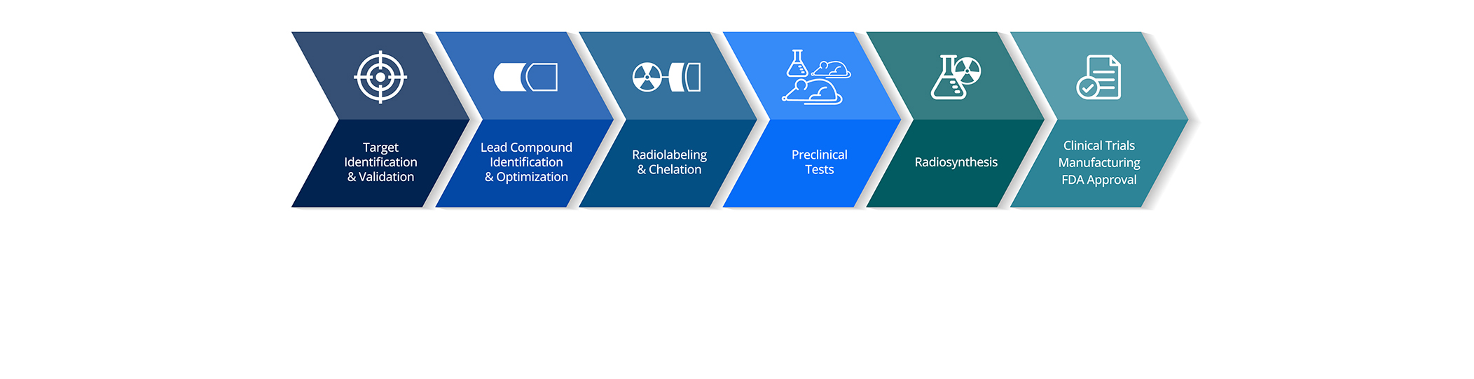
Radiopharmaceutical development brings together some of the most specialized capabilities in oncology research. A single program can require radiochemistry, isotope sourcing, cell-based validation, in vivo model selection, biodistribution, dosimetry, imaging, efficacy assessment, safety interpretation, and translational strategy. Each of these areas requires deep technical expertise, and each produces data that can directly influence whether a program moves forward. What Makes Radiopharmaceutical Development So Complex? The science is complex, but complexity itself is not the biggest risk. The bigger risk is fragmentation. What Is Fragmentation in Radiopharmaceutical Development? Many radiopharmaceutical programs are built across multiple external partners. One group may handle radiochemistry. Another may support in vitro validation. A separate partner may manage animal studies. Imaging may sit elsewhere. Dosimetry may be interpreted by another specialist. Each vendor may deliver high-quality work within their own area, but the full development story can become difficult to connect. What looks like a coordinated program on paper often becomes a collection of parallel workstreams in practice. Why Do Handoffs Create Risk in Radiopharmaceutical Programs? Every handoff introduces risk. A handoff can create delays when materials, data, or protocols need to move between groups. It can create misalignment when one partner’s study design does not fully support the downstream needs of another. It can create data gaps when readouts are generated in different formats, under different assumptions, or without a shared interpretation framework. In radiopharmaceutical development, even small disconnects can affect how confidently a sponsor can interpret results, because the modality relies on tightly linked chemistry, biology, and imaging data. How Can Disconnected Data Affect Radiopharmaceutical Decision-Making? For example, biodistribution data may show tumor uptake, but without a clear connection to target expression or imaging readouts, it may be difficult to determine whether that uptake is biologically meaningful or simply reflective of nonspecific accumulation. Imaging may provide valuable spatial information, but if it is not aligned with tissue collection, dosimetry, or efficacy endpoints, its value can be reduced to a visual reference rather than a decision-support tool. Efficacy may look promising, but without understanding exposure, tumor selectivity, and model biology, the result may not be enough to support a confident development decision or a defensible clinical rationale. What Is the Hidden Cost of Fragmented Radiopharmaceutical Workflows? This is the hidden cost of fragmentation. It is not always visible as a single delay or a single failed experiment. It often appears as uncertainty. Uncertainty can quietly slow internal decision-making. It can lead to repeat studies. It can make it harder to justify dose selection. It can complicate conversations around clinical readiness. It can also make it harder for teams to identify whether a result reflects the agent, the model, the isotope, the study design, or the way the data were generated across separate partners. When a program stalls or produces mixed messages, sponsors often spend significant time trying to reconstruct the reason from disconnected sources. How Fragmentation Increases Program Management Burden Fragmentation also has organizational consequences. When multiple partners are involved, program management overhead increases. Communication becomes more complex. Contract negotiations, material transfers, and regulatory considerations multiply. These are not always visible on a Gantt chart, but they add friction to every decision cycle. For small and mid-sized sponsors in particular, this overhead can absorb resources that would otherwise support scientific progress. What Do Radiopharmaceutical Sponsors Need from an Integrated Workflow? Radiopharmaceutical sponsors need more than isolated technical execution. They need a connected workflow that allows each study to inform the next. Radiochemistry should be planned with downstream in vivo requirements in mind. Model selection should reflect target biology and intended clinical context. Biodistribution and imaging should be designed to answer related questions. Dosimetry should be interpreted alongside tumor uptake, normal tissue exposure, and therapeutic intent. Efficacy should be read as part of a broader translational package, not as a standalone endpoint. An integrated workflow helps create that continuity. How Integrated Radiopharmaceutical Workflows Improve Translational Confidence When radiochemistry, model selection, biodistribution, imaging, dosimetry, and efficacy are aligned within a coordinated study plan, teams can move from data generation to decision-making more efficiently. Results become easier to interpret because the study was designed around the decision the sponsor needs to make. Unexpected findings can be investigated more quickly because the relevant expertise is connected. Data can be reviewed in context rather than assembled after the fact from disconnected sources. Iteration becomes possible, and iteration is often what separates programs that advance from programs that stall. This does not eliminate complexity. Radiopharmaceutical development will always require highly specialized expertise, and no single workflow can remove the intrinsic challenges of the modality. But integration helps ensure that the complexity is managed deliberately rather than passed from one partner to another. It gives sponsors a clearer view of where the risks are and where the strongest evidence is being generated. How to Reduce Risk in Radiopharmaceutical Development For sponsors, the value is not simply operational convenience. It is stronger confidence in the development path. It is fewer surprises during data review. It is clearer alignment with regulators and clinical collaborators. It is a program story that holds together under scrutiny. Fragmentation can make radiopharmaceutical development slower, less predictable, and harder to interpret. Integration can help sponsors reduce handoff risk, preserve data continuity, and make clearer decisions earlier. In a field where the pace of innovation continues to accelerate, the teams that succeed will not only be those with promising science. They will be the teams that can connect the science into a coherent translational strategy, from radiochemistry to clinical translation. See how integrated workflows reduce risk.
Radiopharmaceutical Development: How Strong Preclinical Strategy Builds Translational Confidence
Radiopharmaceutical Development Is Accelerating, and Preclinical Decisions Matter More Than Ever Radiopharmaceutical development is advancing at a pace that would have been difficult to imagine even a few years ago. The 2022 approval of Pluvicto as the first targeted radioligand therapy for PSMA-positive metastatic castration-resistant prostate cancer was a defining moment for the field, helping to validate the clinical potential of radiopharmaceuticals and accelerate broader investment, innovation, and sponsor interest in this modality. New isotopes, targeting vectors, linker chemistries, and therapeutic concepts are expanding what is possible in oncology, and pipelines across biotech and pharma are growing quickly. For drug developers, this is an exciting environment, but it is also one that raises the bar for how programs need to be designed and interpreted. Speed alone is not the central challenge, and the question is whether teams can make strong translational decisions early enough to reduce uncertainty before clinical advancement. A program may move quickly through individual study steps, but if the evidence does not clearly show tumor localization, target engagement, biological activity, and a rational dose strategy, that speed can create risk rather than confidence. In radiopharmaceutical development, a fast preclinical package is only valuable when it is also a defensible one. This is where preclinical strategy becomes critical. Why Preclinical Strategy Is Critical for Radiopharmaceutical Programs A strong preclinical package for a radiopharmaceutical agent needs to answer more than whether the compound can be labeled or whether radioactivity can be measured in tissue. It needs to show whether the agent reaches the tumor, whether uptake is selective, whether target expression aligns with biodistribution, and whether the observed signal translates into meaningful efficacy. These data points are not separate outputs. They are connected pieces of the same translational decision, and they need to be planned together from the start. That connection matters because radiopharmaceutical development introduces layers of complexity that traditional oncology studies do not always capture. The isotope, targeting vector, tumor model, expression profile, biodistribution pattern, dosimetry approach, and therapeutic endpoint all influence how a program should be interpreted. If any one of these elements is considered in isolation, it can create an incomplete or misleading picture. A promising uptake result, for example, is difficult to act on without a clear understanding of target expression, exposure, and downstream biology. Using Clinically Relevant PDX Models to Evaluate Radiopharmaceutical Candidates Clinically relevant PDX models are especially valuable in this environment because they help sponsors evaluate radiopharmaceutical candidates in tumor systems that better reflect human disease. PDX models can capture tumor heterogeneity, target-expression variability, stromal architecture, and treatment-response biology in ways that conventional systems may not fully represent. For radiopharmaceutical programs, that biological context can help determine whether uptake is target-associated, whether tumor selectivity is meaningful, and whether efficacy is likely to translate beyond a narrow experimental setting. When PDX data are combined with biodistribution, imaging, and dosimetry, the translational story becomes far more defensible. Planning Radiopharmaceutical Studies Around PDX Growth, Isotope Availability, and Translational Readiness At the same time, radiopharmaceutical programs need to be honest about what drives their timelines. PDX growth kinetics can be variable, isotope availability can affect study scheduling, radiochemistry optimization can require iteration. These are not obstacles to progress, but are simply realities of the modality that need to be reflected in study planning. Preclinical timelines are not just operational timelines - they are biological and chemical timelines that directly shape how confidently a program can advance toward the clinic. This is where a clinically relevant preclinical platform earns its value. Because PDX models retain the tumor heterogeneity, target expression, and treatment-response biology seen in patients, the data generated early in development carries genuine translational weight, helping sponsors anticipate clinical performance rather than simply satisfying a preclinical checkpoint. The opportunity, then, is not simply to move faster but to build preclinical evidence strong enough to translate directly into clinical strategy. Connecting Biodistribution, Dosimetry, Imaging, and Efficacy in Preclinical Radiopharma Studies That requires early alignment on the right models, endpoints, readouts, and study sequence. Biodistribution and dosimetry data need to be interpreted in the context of target expression and imaging should be connected to downstream efficacy. Efficacy results need to be evaluated alongside exposure, selectivity, and tumor biology. Each step should help teams understand whether the agent is behaving as intended, whether the model is answering the intended question, and whether the program has a clear scientific rationale for continued development. When these elements are planned as part of an integrated preclinical strategy, teams can significantly reduce avoidable uncertainty. They can make more confident go/no-go decisions, refine dose rationale, and identify potential risks before they become more costly at later stages. They can also build a stronger translational narrative for internal stakeholders, investors, and clinical development teams, which is increasingly important in a competitive funding environment. Preparing Radiopharmaceutical Programs for Regulatory, Clinical, and Investor Questions There is also a strategic benefit to preparing for downstream questions early. Regulators, clinical collaborators, and investors will eventually ask how the dose was chosen, why a specific tumor context was prioritized, and how the biodistribution data support the therapeutic index. Programs that plan for these questions during preclinical development are better positioned to answer them with clarity rather than reconstruct the story from disconnected studies later. Building Translational Confidence in Radiopharmaceutical Development Radiopharmaceutical development is moving fast, and that momentum is a positive sign for the field. But speed only creates value when it is supported by evidence that is biologically meaningful, operationally reliable, and translationally connected. For teams advancing radiopharmaceutical programs, the real advantage will come from building confidence early, through preclinical strategies that connect chemistry, biology, imaging, and interpretation into a single decision-making framework. Faster progress is welcome. Better decisions are essential. Build confidence in your next radiopharmaceutical development decision.
How Preclinical PDX Studies Help Guide PSMA Radioligand Development
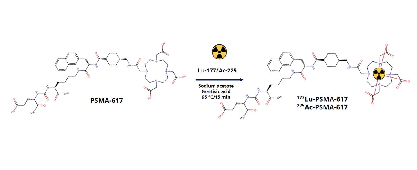
Metastatic castration-resistant prostate cancer, or mCRPC, remains a major clinical challenge. While prostate-specific membrane antigen, PSMA, has emerged as a highly promising therapeutic target, differences in tumor biology, target expression, and radiopharmaceutical behavior continue to influence clinical outcomes. For PSMA-targeted radioligand therapies to reach their full potential, developers need preclinical models that reliably capture both tumor complexity and biodistribution behavior in vivo. At AACR 2026, we presented data showing how patientderived xenograft (PDX) models can be used to evaluate PSMA-targeted radiopharmaceuticals at multiple levels. This included radiochemistry quality, biodistribution, and tumor selectivity. The work focused on two clinically relevant agents, [177Lu]LuPSMA617 and [225Ac]AcPSMA617, tested across a panel of mCRPC PDX models with variable PSMA expression. Why PDX models are critical for radiopharmaceutical development Radiopharmaceutical therapies rely on far more than target binding alone. Successful translation depends on tumor uptake, retention, clearance from healthy tissues, and therapeutic index. Traditional cell line models fail to capture the architectural and microenvironmental features that shape these processes in patients. PDX models preserve the genetic heterogeneity and tissue organization of the original human tumor. This makes them particularly well suited for studying radioligand distribution and efficacy in vivo. In the context of PSMA-targeted therapies, PDX models allow us to directly evaluate how differences in PSMA expression and tumor biology influence radiotracer uptake in clinically relevant tumor models. Designing a clinically relevant PSMA radiopharmaceutical study In this study, we evaluated PSMA617 labeled with either lutetium177 or actinium225 across multiple prostate cancer PDX models representing metastatic, castration-resistant disease. The models spanned a range of PSMA expression levels, enabling us to assess how target expression directly relates to biodistribution. We first confirmed robust radiochemical performance. PSMA617 was efficiently labeled with both isotopes, achieving high radiochemical purity that met stringent quality standards. Establishing radiochemistry consistency is a critical first step before moving into in vivo evaluation, particularly for comparative studies involving different isotopes. How biodistribution studies inform target specificity and safety Following radiotracer administration, we conducted biodistribution analyses at defined time points to quantify uptake across tumors and major organs. Rather than relying on a single model, we assessed multiple PDXs to capture biologically driven variability. Across the panel, tumor uptake correlated strongly with PSMA expression levels. Models with high PSMA expression showed selective and robust radiotracer accumulation, while low-PSMA models demonstrated minimal uptake. This clear relationship confirms biological predictability and reinforces the value of molecularly annotated PDX panels when evaluating PSMA-targeted therapies. Importantly, although both [177Lu]LuPSMA617 and [225Ac]AcPSMA617 demonstrated tumor targeting, differences emerged in tumor-to-normal tissue ratios. These distinctions highlight why side-by-side preclinical evaluation is essential when selecting isotopes and dosing strategies for clinical development. Comparing beta- and alpha-emitting PSMA therapies One of the most important questions in PSMA radiopharmaceutical development is how different isotopes behave in vivo. Lutetium177 and actinium225 differ in emission properties, tissue penetration, and potential toxicity profiles. Our comparative biodistribution analyses showed that while both agents localize to PSMA-expressing tumors, [177Lu]LuPSMA617 demonstrated more favorable tumortotissue ratios across several organs. These findings provide early insight into how isotope choice may influence therapeutic window and tolerability. While alpha-emitters remain highly compelling for their potent cytotoxicity, data like this emphasize the importance of rigorous preclinical evaluation in clinically relevant models before advancing treatment strategies. Why this approach matters for radiopharmaceutical pipelines Radiopharmaceutical development is inherently multidisciplinary, integrating chemistry, biology, imaging, and therapeutic evaluation. PDX models provide a unifying platform where all of these components can be assessed in context. Using well-characterized mCRPC PDXs allows us to: Link PSMA expression directly to in vivo uptake Compare isotopes in clinically relevant tumor models Evaluate tumor selectivity alongside therapeutic response Reduce translational uncertainty before clinical studies This kind of integrated preclinical strategy is especially valuable as PSMA-targeted therapies continue to expand into earlier disease settings and combination regimens. Want to see the full dataset? This blog highlights key findings, but the full study includes detailed radiochemistry validation, biodistribution data, tumor-to-tissue ratios, and efficacy results across multiple PDX models. Download our AACR 2026 poster to explore the complete results and analyses.